The Skeletal Structure: Your Hand's Framework
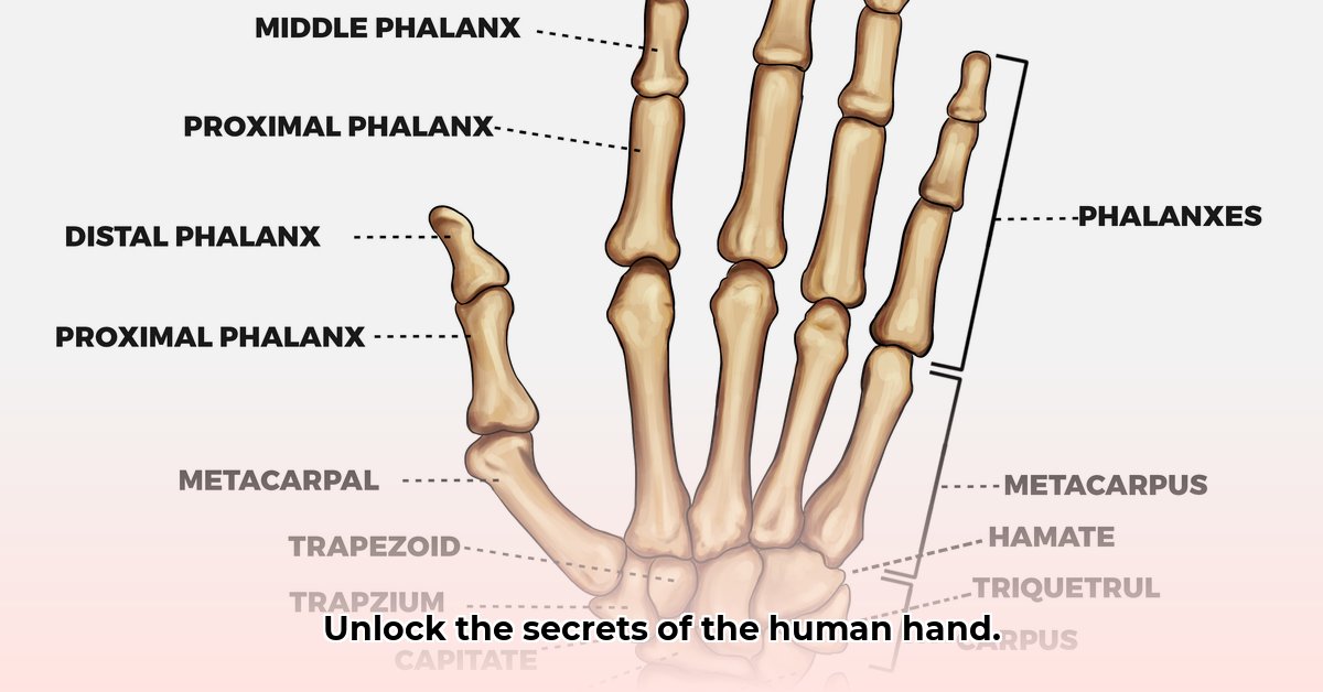
Your hand's bony structure, like a sophisticated scaffolding, supports its complex functions. This framework comprises three key sections: the carpals, metacarpals, and phalanges. The eight small carpals (wrist bones) interlock, enabling flexion, extension, and rotation. Imagine them as cleverly designed building blocks allowing for a surprising range of motion. These provide a stable base for the hand's more distal structures. The five metacarpals form the palm, radiating outwards like spokes from the wrist, each connecting to a finger. Finally, the phalanges are the finger bones; each finger (except the thumb) has three: proximal, middle, and distal. The thumb's unique two-phalange structure is key to our opposable thumb, crucial for grip and dexterity. These bones articulate via joints, enabling an extensive array of movements. Did you know that the intricate arrangement of these bones allows for over 20 different hand positions? For a detailed look at hand anatomy, see this helpful resource: hand anatomy.
The Muscular System: The Power Behind the Precision
The muscles of the hand are divided into two groups: intrinsic and extrinsic. The intrinsic muscles, residing entirely within the hand, are responsible for fine motor control. These tiny but powerful muscles allow for precise movements like writing or buttoning a shirt. In contrast, the extrinsic muscles originate in the forearm, providing the power for stronger grips needed for tasks such as carrying heavy objects or using tools; they act as the powerhouse complementing the precision of the intrinsic muscles. Multiple muscle groups coordinate to achieve flexion, extension, adduction, and abduction of the fingers—a complex interplay enabling our remarkable hand dexterity. How many individual muscles do you think contribute to the movement of your thumb alone? (There are actually nine!)
The Ligaments: Keeping Everything Together
The hand's bones are intricately connected and stabilized by ligaments—strong fibrous bands acting like incredibly strong ropes. These ligaments are essential for maintaining the hand's structural integrity and preventing injuries. Key ligaments include the collateral ligaments, preventing excessive sideways bending of fingers, and the volar plates, preventing backward bending. The palmar fascia, a thick band of tissue in the palm, adds further support and structure. This complex network ensures the stability and resilience of the hand. Think about the stresses placed on your hand daily; it's a testament to the strength and design of this ligamentous support system.
The Nervous System & Blood Vessels: The Communication Network
The hand's remarkable dexterity requires a sophisticated nervous and vascular system. Three main nerves—median, ulnar, and radial—transmit sensory information and motor commands between the brain and the hand. These act like communication superhighways, transmitting information crucial for both sensation and movement. Damage to any of these nerves can dramatically affect hand function and sensation. Meanwhile, a rich arterial system supplies oxygenated blood, while a complementary venous system removes waste products. This constant blood flow is vital for maintaining the hand’s health and function. Dr. Anya Sharma, Hand Surgeon at Massachusetts General Hospital, states, "The intricate interplay between nerves and blood vessels is crucial for maintaining the hand's vitality and functionality. Compromise in either can have significant impacts on dexterity and overall hand health."
Hand Function: Power and Precision
The hand's versatility is apparent in its ability to perform both power and precision grips. Power grips, utilizing the entire hand, are suited for tasks like hammering or carrying heavy bags. Conversely, precision grips, employing mainly the fingers and thumb, enable intricate actions like writing or picking up small objects. This adaptable design highlights the hand's exceptional engineering. This dual functionality is a testament to the coordinated actions of bones, muscles, ligaments, and nerves working in perfect harmony. How many everyday tasks rely on this unique combination of power and precision? Hundreds, if not thousands!
Clinical Significance: Understanding Hand Injuries and Conditions
A thorough knowledge of hand anatomy is paramount for diagnosing and treating various hand injuries and conditions. For medical professionals, precise localization of bones, muscles, nerves, and ligaments is crucial for effective diagnosis and treatment planning. This knowledge is fundamental to managing conditions such as carpal tunnel syndrome, tendon injuries, fractures, and arthritis, showcasing the critical link between anatomical understanding and successful clinical practice. Understanding the anatomy of the hand, in essence, is understanding the foundation of successful hand surgery and rehabilitation.
How to Compare Muscle Fiber Arrangement in Thenar Muscles Using Anatomical Atlases
This section details a method for comparing muscle fiber arrangement in thenar muscles (abductor pollicis brevis, flexor pollicis brevis, opponens pollicis) using anatomical atlases. The process involves analyzing detailed illustrations from multiple angles, noting variations in fiber orientation, and correlating findings with muscle function. This detailed approach enhances our understanding of these muscles’ role in thumb movement.
Key Takeaways:
- Analyzing thenar muscles requires going beyond simple median sections to understand their complexity.
- Anatomical atlases provide detailed views necessary for comparing fiber arrangement.
- Comparing multiple atlases reveals variations and patterns in fiber orientation. This analysis significantly improves the understanding of thenar muscle function and its implications for hand movement.
Steps for Comparison:
- Choose reputable atlases: Select atlases known for accurate and detailed illustrations (e.g., Gray's Anatomy).
- Use multiple views: Analyze illustrations from various perspectives (anterior, posterior, lateral) to gain a comprehensive understanding.
- Compare and contrast: Note any differences in fiber direction, density, and fascicle organization across different atlases.
- Consider muscle layers: Analyze fiber orientation within each layer of the thenar muscles for a more detailed understanding.
- Correlate with function: Relate your observations on fiber arrangement to the known actions of the thenar muscles and how this arrangement contributes to thumb movement precision and strength.
Remember individual variations exist, and the methods used to prepare specimens for illustration can influence results. By combining visual observation with an understanding of muscle function, one can gain a rich understanding of thenar muscle architecture, with implications for both clinical practice and research.